FlowCam customers frequently ask us how to optimize analysis of Microcystis, a globally pervasive colonial cyanobacteria (blue-green algae) which is seen in source water systems and recreational lakes. The colonies can range in size from a few microns up to several thousand microns in diameter. Customers are concerned about losing part of the sample or not being able to capture a quality image due to the colony’s size and density. Dense colonies superimpose cells in the image, making it easy to underestimate cell counts, while large colonies can make it challenging to determine what objective is best used to image the sample.
Question: What protocol does Fluid Imaging recommend to analyze colonies and scums of Microcystis?
Answer:
- There are multiple methods to process a Microcystis sample. Methods will vary depending on the density and concentration of the colonies in the sample, but troubleshooting the best solution is taught during FlowCam training. The following peer reviewed procedure that was developed by the California Department of Water Resources (USA) is commonly used to count cells of colonial cyanobacteria, including Microcystis and Anabaena. This strategy parallels that of the microscope, but can leverage a larger and more statistically significant data set. Click on this link to review the method. Cell Enumeration of Colonial and Filamentous Cyanobacteria with FlowCam: Method & Case Study
- The flow chamber is 100µm deep for the 10X objective and 300 or 600µm deep for the 4X objective. The 10X objective is most commonly used, but the operator can select a larger flow cell if needed.
- Microcystis will change size/shape according to the environment that it’s in. It’s common to see a 300µm colony pass through a 100µm deep flow cell. Dilution with de-ionized water can also facilitate smoother processing if the colonies are large, ensuring that the colonies are dispersed enough to pass through the flow chamber one at a time. Microcystis is more forgiving than filamentous organisms like Aphanazomenon, which are more likely to clog. Then again, Aphanazomenon sometimes only exhibits as a single filament, in which case, it’s unlikely to clog. (As usual with biological sciences… “it depends”.)
- Samples are often (but not always) filtered with mesh to remove large sediments/colonies to eliminate clog potential. If a clog occurs in the new FlowCam Cyano or FlowCam 8000, it takes 2 minutes to address. A clog in our older model, the FlowCam VS can take up to 15 minutes to resolve or require a new flow cell. If the sample is filtered with 100µm mesh for the 100µm flow cell then you can do one of two things:
- Analyze a second sample at 4X with a larger flow cell (300 or 600µm deep).
- Rinse the mesh onto a slide and briefly scan it under a microscope to visually confirm if the data resembles that which is quantified by the FlowCam. This is common since the microscope is an excellent tool for qualitative (as opposed to quantitative) data collection and requires little additional time for the operator.
- In the case of a bloom, the operator can sonicate/shake the sample to disperse the colonies so the cells can be counted individually.
- Cell counts are not an indication of toxicity; instead they are a low cost, fast procedure used to quantify risk. Toxin tests can become extremely expensive, so it’s cost and time efficient to identify if toxin producing organisms are present and significantly abundant. Click here for more information:
Use an Integrated Approach to Monitor Algal Blooms
We also love to hear from our FlowCam users. What methods and strategies have you employed in these situations?
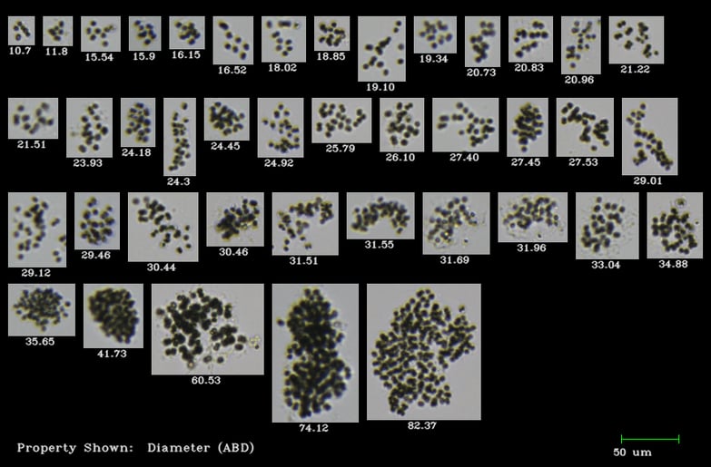
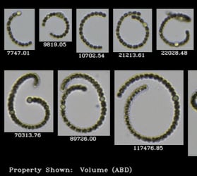
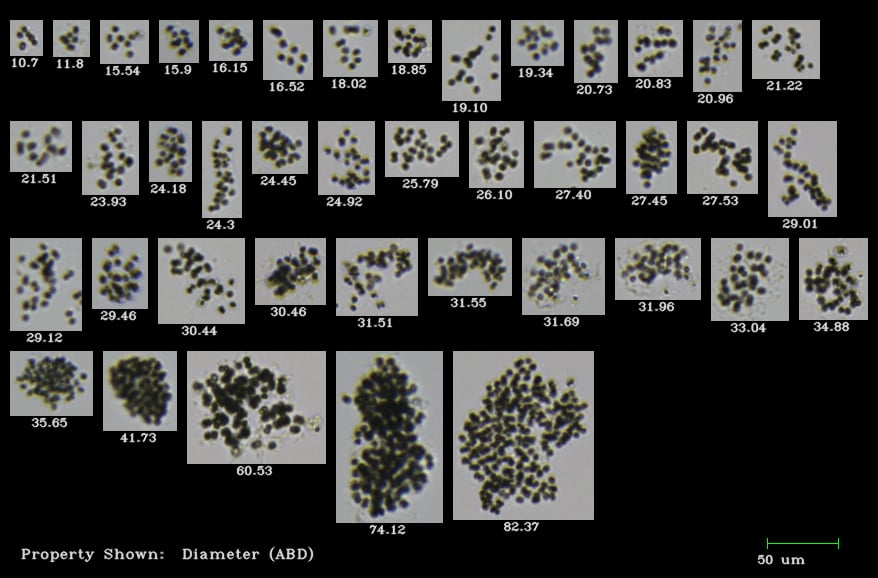
Pictured below: Microcystis Colonies imaged by the FlowCam (image labels show colony diameter in microns)